


Ekaterinburg, Ekaterinburg, Russian Federation
Ufa, Ufa, Russian Federation
from 01.01.1981 until now
Ufa, Ufa, Russian Federation
Background. Periodontal disease is one of the most common and complex pathologies in dentistry. It is known frequently damage to the tissues of the periodontal complex with dermatoses. The most relevant among the dermatoses of the mucous membrane of the mouth and the red border of the lips is oral lichen planus. Among the six clinical forms of red flat oral lichen planus and the red border of the lips, the exudative-hyperemic and erosive-ulcerous forms occur most often. Atypical form is much less common than other forms and is often diagnosed by dentists as an inflammatory periodontal disease. At the same time, the pathogenetic mechanisms of inflammatory processes in the gums, which are different in oral lichen planus, are not taken into account, which, accordingly, complicates adequate treatment. Objectives. The aim of the study was to analyze the periodontal status in patients with exudative-hyperemic, erosive-ulcerative and atypical forms of oral lichen planus. Methods. Under our supervision there were 181 patients with oral lichen planus, in whom a simplified Green-Vermillion hygiene index was determined. To assess the state of periodontal used periodontal index according to Russell. Results. When assessing the hygienic condition of the oral cavity, high values were observed in individuals with severe forms of the disease: erosive-ulcerative, exudative-hyperemic. The highest values of the periodontal index were found in patients with atypical, erosive and ulcerative forms of oral lichen planus. Conclusions. The results of the clinical examination of patients with oral lichen planus dictate, first of all, to include in the scheme of complex treatment of it the sanitization of oral cavity and complex treatment of inflammatory periodontal diseases as well.
oral lichen planus, exudative-hyperemic form, erosive-ulcerative form, atypical form, periodontal index
Введение
Заболевания пародонта представляют одну из наиболее распространенных и сложных патологий в стоматологии, они занимают второе место после кариеса [1, 2].
Распространенность этого заболевания среди взрослого населения остается на высоком уровне и не имеет тенденции к снижению [3, 4]. Особенно остро встает вопрос о патологии пародонта у лиц с сочетанной патологией в связи с возможным взаимоотягчающим характером течения [5―7]. Установлено, что степень нарушений пародонтального статуса и гигиены полости рта, например, при пузырчатке, во многом определяет агрессивность клинического течения [8]. Известна высокая частота поражения тканей пародонтального комплекса при дерматозах [9, 10]. По данным отечественных и зарубежных специалистов [11―14], частота выявления специфических заболеваний пародонта при красном плоском лишае полости рта варьируется от 13,0 до 48,0 % [15,16] (рис.1).
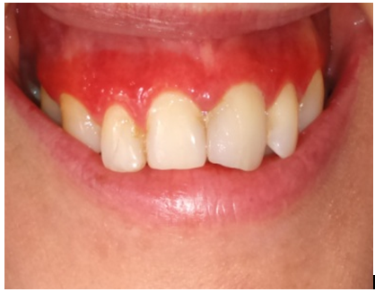
Рис.1. Пациентка Л., 42 года. КПЛ СОР, атипичная форма
Fig.1. Patient L., 42 years old. OLP, atypical form
Красный плоский лишай (КПЛ) ― хронический, длительно текущий дерматоз мультифакторной природы с многообразными клиническими проявлениями и вовлечением в процесс кожи, ее придатков (волос, ногтей) и слизистых оболочек [17―24]. Его локализация может быть различной, наиболее часто встречается на слизистой оболочке (СО) щек, языка, а также десне [25, 26, 29].
Согласно классификации, предложенной Машкиллейсоном А.Л. и Боровским Е.В. (1984), выделены следующие клинические формы КПЛ СОР: типичная, экссудативно-гиперемическая, эрозивно-язвенная, гиперкератотическая, атипичная, буллезная.
Среди шести клинических форм КПЛ слизистой оболочки рта и красной каймы губ наиболее часто встречаются и тяжело протекают экссудативно-гиперемическая и эрозивно-язвенная. Непрерывно рецидивирующее течение данных форм затрудняет проведение гигиены полости рта, что, в свою очередь, отягощает клиническое течение пародонтита [27].
Атипичная форма встречается значительно реже остальных и зачастую диагностируется врачами-стоматологами как воспалительное заболевание пародонта (рис. 2, 3).
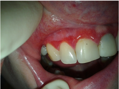
Рис.2. Пациентка А., 43 года. КПЛ СОР, атипичная форма
Fig.2. Patient A., 43 years old. OLP, atypical form
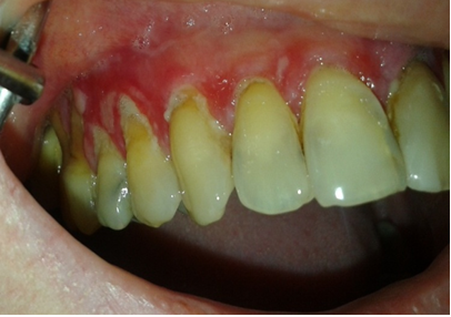
Рис. 3. Пациентка К., 46 лет. КПЛ СОР, атипичная форма
Fig. 3. Patient K., 46 years old. OLP, atypical form
При этом не учитываются патогенетические механизмы воспалительных процессов в десне, которые отличаются при КПЛ СОР, что, соответственно, затрудняет адекватное лечение. Имеется ряд механизмов при КПЛ СОР, определяющих воспаление в пародонте: иммунный дисбаланс, нарушения микроциркуляции, дефицит микроэлементов. Воспалительно-деструктивные изменения пародонта, в свою очередь, способствуют эндотоксемии и становятся фактором риска развития рецидивов КПЛ СОР [28―31].
В связи с вышеизложенным целью исследования явилось изучение пародонтального статуса у больных с экссудативно-гиперемической, эрозивно-язвенной и атипичной формами КПЛ СОР.
Материал и методы
Под нашим наблюдением находились 181 пациент в возрасте от 24 до 76 лет с различными формами КПЛ, обратившихся за консультативной помощью в клиники стоматологии при УГМУ и БГМУ. Для оценки гигиенического состояния полости рта пациентов использовали упрощенный индекс гигиены по Грину ― Вермиллиону (OHI-S), для оценки состояния пародонта ― пародонтальный индекс (ПИ) по Расселу. Статистическая обработка данных выполнялась с помощью методов медико-биологической статистики в пакете Statistica.
Критериями включения пациентов в исследование были:
наличие клинических проявлений красного плоского лишая слизистой оболочки рта, позволяющих поставить диагноз «красный плоский лишай (L43)» в соответствии с МКБ-10 (1997);
наличие информированного согласия на участие в исследовании.
Критерии исключения пациентов:
отсутствие клинических проявлений красного плоского лишая слизистой оболочки рта, позволяющих поставить диагноз «красный плоский лишай (L43)»;
наличие соматической патологии в стадии декомпенсации;
наличие онкологических заболеваний;
несоблюдение протокола исследования, отказ больного от проведения исследования;
беременность, лактация;
выявление атипичных клеток при гистологическом исследовании СОР.
Результаты и обсуждение
У 67 пациентов выявлена экссудативно-гиперемическая форма. Упрощенный индекс гигиены OHI-S в исследуемой группе составил 2,9±1,1. Поражение тканей пародонта выявлено у 32 пациентов, которые проявлялись в виде хронического генерализованного пародонтита средней степени тяжести, где индекс ПИ был равен 3,95±1,01.
Эрозивно-язвенная форма ― самая тяжелая из всех ― выявлена у 75 пациентов. Индекс гигиены в данной группе составил 3,65±1,12 балла, что значительно ниже, чем при других формах.
Патология тканей пародонта выявлена у 35 больных, индекс ПИ был равен 3,98±0,21, что соответствует пародонтиту средней и тяжелой степеней тяжести.
У 39 пациентов выявлена атипичная форма, которая возникала на слизистой оболочке верхней губы и на соприкасающейся с ней десне. Упрощенный индекс гигиены OHI-S в исследуемой группе составил 3,5±0,31.
Поражения тканей пародонта выявлены у всех пациентов (100 %), которые проявлялись в виде хронического генерализованного пародонтита средней степени тяжести, где индекс ПИ был равен 2,65±0,45.
Таким образом, при оценке гигиенического состояния полости рта с помощью упрощенного индекса ОHI-S высокие значения были у лиц с тяжелыми формами заболевания: эрозивно-язвенной и экссудативно-гиперемической.
Наиболее высокие значения индекса ПИ выявлены у больных с атипичной и эрозивно-язвенной формами КПЛ СОР, что соответствовало пародонтиту тяжелой и средней степеней тяжести.
Полученные результаты клинического обследования пациентов могут подтверждаться рядом взаимосвязанных факторов: невозможностью проведения гигиены полости рта на фоне воспаления и боли из-за наличия очагов поражения на СОР, усилением раздражения от применяемых средств гигиены, нарушением процесса очищения в результате изменения физико-химических свойств ротовой жидкости, развитием дисбаланса микробной флоры. Следствием вышеперечисленных факторов может быть нарушение гомеостаза в полости рта, усугубляющее течение КПЛ СОР, прогресс воспалительных заболеваний пародонта, что диктует, в первую очередь, включать в схему комплексного лечения КПЛ СОР санацию полости рта, а также комплексное лечение воспалительных заболеваний пародонта.
1. Yanushevich, O. O., Dmitreva, L. A., Revazova, Z. E. (2012). Parodontit XXI vek [Parodontitis XXI century]. Moscow : MSMSU, 366. (In Russ.)
2. Yefimova, O. V., Ushnitskiy, I. D., Sozonov, I. G. (2008). Sovremennyye aspekty vospalitel'nykh zabolevaniy parodonta [Modern aspects of inflammatory periodontal diseases]. Yakutskiy meditsinskiy zhurnal [Yakutsk Medical Journal], 24, 4, 77-80. (In Russ.)
3. Iordanishvili, A. K., Tikhonov, A. V., Ar'yev, A. L. (2010). Vozrastnaya epidemiologiya zabolevaniy parodonta [Age-related epidemiology of periodontal diseases]. Parodontologiya [Parodontologiya], 15, 1 (54), 25-28. (In Russ.)
4. Yanushevich, O. O., Kuz'mina, I. N. (2009). Sostoyaniye tkaney parodonta u naseleniya v vozraste 35-44 let v regionakh Rossii [The condition of periodontal tissues among the population aged 35-44 years in the regions of Russia]. Rossiyskiy stomatologicheskiy zhurnal [Russian dental journal], 1, 43-45. (In Russ.)
5. Kiran, M., Arpak, N., Unsal, E. et al. (2005). The effect of improved periodontal health on metabolic control in type 2 diabetes mellitus. J Clin Periodontol, 32, 3, 266-272. DOI :https://doi.org/10.1111/j.1600-051X.2005.00658.x
6. Mustapha, I. Z., Debrey, S., Oladubu, M. et al. (2007). Markers of systemic bacterial exposure in periodontal disease and cardiovascular disease risk: a systematic review and meta-analysis. J Periodontol, 78, 12, 2289-2302.
7. Wilder, R. S., Iacopino, A. M., Feldman, C. A. et al. (2009). Periodontal-systemic disease education in U.S. and Canadian dental schools. J Dent Educ, 73, 1, 38-52.
8. Arduino, P. G., Farci, V., D’Aiuto, F. et al. (2011). Periodontal status in oral mucous membrane pemphigoid: initial results of a case-control study. Oral Diseases, 17 (1), 90-94.
9. (2015). Kaur Supreet. Periodontal manifestations of dermatitis herpetiformis: a case report. Indian j of Comprehensive Dental Care, 2, 643-646.
10. Holmstrup, P., Schiotz, A. W., Westergaard, J. (1990). Effect of dental plaque control on gingival lichen planus. Oral Surg Oral Med Oral Pathol, 69, 5, 585-590.
11. Ivanova, Ye. V., Shcherbakova, E. G., Rabinovich, O. F. (2005). Sovremennyye podkhody k patogeneticheskoy terapii ploskogo lishaya slizistoy obolochki polosti rta [Modern approaches to pathogenetic treatment of lichen planus of the oral mucosa]. Stomatologiya [Dentistry], 5, 31-34. (In Russ.)
12. Gileva, O., Libik, T., Khalilayeva, E., Gulyaeva, Y., Khaliavina, I., Podgornii, R. (2008). Oral health related quality of life in patients with non-specific ulcero-necrotic oral mucosal lesions. Oral Diseases, 14, l, 24.
13. Axell, T., Rundquist, L. (1987). Oral lichen planus a demographic study. Community Dent. Oral. Epidemiol, 15, 1, 52-56.
14. Sharma, S., Saimbi, C. S., Koirala, B. (2008). Erosive oral lichen planus and its management: a case series. JNMA, 47, 2, 86-90.
15. Ertugrul, A. S., Arslan, U., Dursun, R., Hakki, S. S. (2013). Periodontopathogen profile of healthy and oral lichen planus patients with gingivitis or periodontitis. International journal of oral science, 5 (2), 92-97.
16. Libik, T. V., Gileva, O. S., Bondarenko, Ye. A., Beleva, N. S. (2008). Kompleksnaya otsenka vliyaniya zubnykh past na slizistuyu obolochku polosti rta i krasnuyu kaymu gub (Chast' I) [Comprehensive assessment of the influence of toothpastes on the mucous membrane oral cavities and red lip rim (Part I)]. Institut stomatologii [Institute of Dentistry], 1 (38), 26-28. (In Russ.)
17. Canto, A. M., Muller, H., Freitas, R. R., Santos, P. S. (2010). Oral liehen (OLP): clinical and complementary diagnosis. Anais Brasileiros de Dermatologia, 85 (5), 669-675.
18. Usatine, R. P., Tinitigan, M. (2011). Diagnosis and Treatment of Lichen Planus. Am. Fam. Physician, 84, 1, 53-60.
19. Thompson, L. D. (2012). Oral lichen planus. Ear Nose Throat J. 91, 3, 102-104.
20. Shekar, C., Ganesan, S. (2011). Oral lichen planus: Review. J. Dent. Sci. Res, 2, 1, 62-87.
21. Gileva O. S. (2015). Zabolevaniya slizistoy obolochki polosti rta: osnovnyye trendy v sovremennoy stomatologii [Diseases of the oral mucosa: the main trends in modern dentistry]. Maestro stomatologii [Maestro dentistry], 4 (60), 17-22. (In Russ.)
22. Chuykin, S. V., Akmalova, G. M., Chernysheva, N. D. (2015). Osobennosti klinicheskogo techeniya krasnogo ploskogo lishaya slizistoy obolochki rta, assotsiirovannogo s gerpesvirusnoy infektsiyey [Features of the clinical course of lichen planus of the oral mucosa associated with herpes virus infection]. Sovremennyye problemy nauki i obrazovaniya [Modern problems of science and education], 2, 80. (In Russ.)
23. Yegorova, Ye. G., Kudashkina, N. V., Akmalova, G. M. (2015). Fitopreparaty v kompleksnom lechenii zabolevaniy slizistoy obolochki rta [Phytodrugs in the complex treatment of diseases of the oral mucosa]. Mezhdunarodnyy zhurnal prikladnykh i fundamental'nykh issledovaniy [International Journal of Applied and Basic Research], 12-7, 1223-1227.
24. Chuykin, S. V., Akmalova, G. M., Chuykin, O. S., Makusheva, N. V., Akatyeva, G. G. (2016). The role of mineral elements in the pathogenesis of lichen planus of the oral mucosa. Research Journal of Pharmaceutical, Biological and Chemical Sciences, 7, 6, 704-710.
25. Lo Russo, L., Guiglia, R., Pizzo, G. et al. (2010). Effect of desquamative gingivitis on periodontal status: a pilot study. Oral Diseases, 16 (1), 102-107.
26. Scully, C., Carrozzo, M. (2008). Oral mucosal disease: lichen planus. British Journal of Oral and Maxillofacial Surgery, 46 (1), 15-21.
27. Azizi, A., Rezaee, M. (2012). Comparison of periodontal status in gingival oral lichen planus patients and healthy subjects. Dermatology research and practice, 561-562.
28. Ertugrul, A. S., Arslan, U., Dursun, R., Hakki, S. S. (2013). Periodontopathogen profile of healthy and oral lichen planus patients with gingivitis or periodontitis. International journal of oral science, 5 (2), 92-97.
29. Scattarella, A., Petruzzi, M., Ballini, A. et al. (2011). Oral lichen planus and dental hygiene: a case report. 9, 2, 163-166.
30. Thompson, L. D. (2012). Oral lichen planus. Ear Nose Throat J, 91, 3, 102-104.
31. Shekar, C. (2011). Oral lichen planus: Review. J. Dent. Sci. Res, 2, 1, 62-87.
